Ion Exchange of Proteins with Organic Solvents
Organic solvents “denature” proteins; that is, they cause changes in tertiary structure that may cause enzymes and other proteins to lose biological activity. However, many proteins will regain their activity once the organic solvent is removed. Also, in proteomics analyses, the objective is not to isolate a biologically active protein but just to see how much of which proteins are present. In ion-exchange chromatography (IEX) of proteins, adding organic solvent to the mobile phases can sometimes improve results dramatically.
The following example shows IEX of recombinant α-bungarotoxin on a PolyCAT™ A column. The protein was expressed in the yeast strain P. pastoris. This choice was unfortunate, since P. pastoris tends to attach glycan side chains to proteins that normally don’t have them, as in this case. The resulting mixture of glycoforms eluted in an envelope with a regular salt gradient [TOP]. When 40% ACN was included in the mobile phases, though, then the various forms were well-resolved [BOTTOM]. For many proteins like this in the range 15-30 KDa (growth factors, etc.), a range of 30-40% organic solvent yields optimal separations. Order PolyCAT A™ now.

If a protein isn’t normally found in an aqueous medium, such as a membrane protein, it may not be possible to do IEX at all without some organic solvent. The example below concerns Lung Surfactant Protein in an emulsion with 500 parts lipid. With 70% organic solvent in the mobile phase, hydrophilic interaction is superimposed on top of the electrostatic effects. Under these conditions, lipid is not retained and elutes in the void volume. The protein is then eluted with a salt gradient. A number of variants are evident, corresponding to truncation sequences with deletions from either terminus.
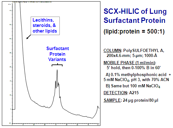
– data courtesy of Richard Hartwick (PharmAssist) –
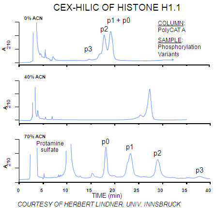
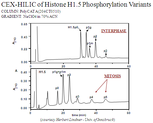
Hydrophilic interaction becomes significant if the mobile phase is > 60% organic, as in the above example. These HILIC effects are superimposed on the electrostatic effects. In the following example, phosphorylation variants of Histone H1 are separated on a PolyCAT A™ column. Histones are basic proteins and are well-retained on a cation-exchange column. With 0% ACN in the mobile phases [TOP], the more extensively phosphorylated variants elute early, since the negative charge of the phosphate repels the negatively-charged stationary phase. Overall, resolution is poor. With 70% ACN in the mobile phases [BOTTOM], resolution is excellent, with the order of elution inverted. Under these conditions the hydrophilic interaction with a phosphate group is greater than its electrostatic repulsion, leading to a net increase in retention. With 40% ACN [MIDDLE], both forces are in balance, and all variant forms coelute.
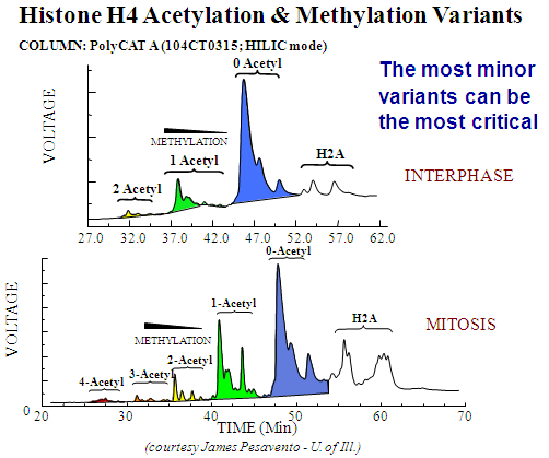
Histone H4 acetylation and methylation variants: A cation-exchange column can separate variants with acetylated Lys- residues, since they vary in the number of + charges. However, with hydrophilic interaction superimposed on the electrostatic effects, the column can also separate variants with the same number of acetylated Lys- residues but differing in the number of methylated Lys- residues. In the figure below, the more highly acetylated forms are present in the smallest amounts and for the shortest time in the cell cycle, but their presence is required if the cell is to enter mitosis. Their identification was only possible by adding this top-down separation step prior to digestion and bottom-up peptide identification. Otherwise, they would be masked by the far more abundant, less highly acetylated variants.
Histone H1.5 phosphorylation variants: HPLC is superior to electrophoresis for separation of protein variants. The figure below shows the isolation of Histone H1.5 and the separation of phosphorylation variants by HPCE.
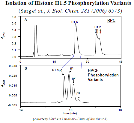
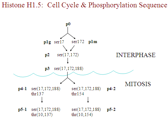
When the same mixture is resolved by cation-exchange in the HILIC mode [BELOW], the resulting peaks are more than twice as sharp. Furthermore, the column is able to distinguish between positional variants, each bearing a single phosphate group but on different Ser- residues. When the cell is in mitosis, more highly phosphorylated forms appear that are absent in interphase. Again, these forms are required for successful entry of the cell into mitosis. The schematic below shows the sequence of phosphorylation. The variants with 4 or 5 phosphates represent the first observation of a histone phosphorylated at a Thr- residue. Again, these low-abundance forms could only be identified by first separating them prior to digestion from more abundant variants.
PolyCAT A™ and PolySULFOETHYL A™ are trademarks of PolyLC Inc. All Rights Reserved